

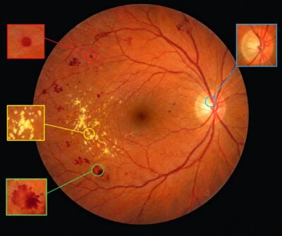
Deep-learning analysis of color fundus images to support retinal screening workflows
| APPLICATION Ophthalmic imaging / diabetic eye screening |
INPUT Color retinal fundus photographs |
OUTPUT Image-quality assessment, automated grading support and lesion heatmap |
The Challenge
Diabetic retinopathy is a diabetes-related retinal condition that can threaten vision. Color fundus photography provides a practical way to document the retina, but screening workflows must handle large numbers of images and differences in image quality. Images that are blurred or do not clearly show the macula and retinal vessels can limit reliable assessment. A consistent first-pass analysis can help prioritize review and support screening teams.
The Imaging and AI Approach
ADCIS describes an automatic diabetic-retinopathy grading application that analyses color fundus images using deep-learning algorithms. The software includes a real-time image-quality assessment and can be deployed as a cloud-based solution, a stand-alone application, or embedded within ophthalmic equipment. ADCIS states that the application is compatible with color fundus cameras available on the market.
The quality assessment considers visibility of the macula and peripheral vessels, overall image sharpness, visibility of vessel boundaries and vessel density. The grading workflow then analyses the fundus image and can display a heatmap highlighting pixels considered most significant to the algorithm’s retinal-lesion assessment. Such a heatmap offers a visual aid for reviewing the areas that contributed to the software output; it should not be interpreted as a stand-alone diagnosis.
Fundus Image: Retinal Structures and Representative Findings
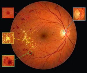
Illustrative fundus image created for this case study—not an ADCIS product screenshot, patient image, or validated clinical annotation.
The illustration indicates representative features commonly discussed in diabetic-retinopathy image analysis: small red lesions such as microaneurysms, retinal hemorrhages, bright hard exudates, retinal blood vessels and the optic disc. The macular region is the darker area near the centre of the illustration. The precise presence, type and clinical significance of any lesion must be established through validated image analysis and qualified clinical assessment.
Reported Performance
On the ADCIS application page, the company reports image interpretability of 96.4%, sensitivity of up to 99% and specificity of 87%. It also reports a processing time of approximately 0.4 seconds per image on a standard CPU. ADCIS’s accompanying note qualifies the sensitivity figures by referencing different partner configurations and a false-negative adjustment for one result. These figures are vendor-reported and should be read in the context of the company’s stated evaluation conditions; they are not a guarantee of performance for every camera, population, clinical setting or deployment.
Operational Benefits
Potential Workflow
Capture a color fundus image → assess image quality → analyse the image with the grading algorithm → review the output and lesion heatmap → refer for clinician assessment or further examination as required.
Source and Acknowledgement
This case study is based on the application description published by ADCIS: “Automatic Grading of Diabetic Retinopathy.” ADCIS develops image-understanding and image-processing software. The described application, capabilities and performance figures are attributed to ADCIS.
Source: https://www.adcis.net/en/applications/automatic-grading-diabetic-retinopathy/
Disclaimer
This document is an informational application case study prepared from publicly available product information. It is not medical advice and does not establish clinical efficacy, regulatory clearance, or suitability for a specific intended use. The fundus illustration is synthetic and is included only to explain representative retinal structures and lesion types; it is not a real patient image and is not the output of ADCIS software. Automated image analysis is intended to support—not replace—qualified clinical judgement. Clinical use requires appropriate validation, regulatory and quality-system review, and adherence to the software provider’s current instructions for use. Performance may vary by dataset, camera, image quality, patient population and deployment conditions.
We'll be glad to help you! Please contact our Sales Team for more information.
We'll be glad to help you! Please contact our Sales Team for more information.