

Adjustable zoom optics for observing fast, fine-scale scientific phenomena
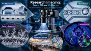
Conceptual illustration for this case study; images are illustrative and are not experimental data or reproductions from the cited papers.
1. Case Study Overview
Scientific research often depends on seeing small structures or rapid events clearly while retaining enough flexibility to adjust magnification, field of view and working distance. Published experimental studies illustrate how adjustable zoom microscope optics can be integrated with high-speed or digital cameras in custom-built imaging rigs. Across fluid dynamics, microfluidics, materials and battery research, and neuroscience imaging, the optical system is selected as part of the experiment—not as a stand-alone lens—so that the target phenomenon can be resolved, recorded and measured.
2. Why Adjustable Zoom Optics Are Used
3. Examples Across Research Disciplines
Fluid Dynamics: Rapid Droplet Spreading
In the study “Short time dynamics of viscous drop spreading,” researchers used synchronized high-speed views to study the early spreading of a viscous drop. The side-view setup paired a high-speed camera with a long-range 12× zoom microscope lens and a 2× adapter tube; the paper reports imaging at 10,000 frames per second and a spatial sampling of 3.5 μm per pixel for that view. This illustrates how zoom optics can help capture a small, fast-moving contact-line region while retaining a practical working distance.
Microfluidics and Chemical Analysis: Droplet Injection
A Scientific Reports study on combining a nanoliter-scale droplet array with high-speed capillary electrophoresis used a high-speed CCD camera and an adjustable microscope zoom lens to record the spontaneous injection process at 4,000 frames per second. The recorded sequence helped researchers examine the thinning and breakup of the liquid bridge between a capillary tip and a droplet—an event difficult to inspect reliably with ordinary-speed imaging.
Microscale Flow: Cavitation in Microchannels
A published study of cavitation in micro-Venturi channels reports an ultra-high-speed camera fitted with a 12× zoom lens and optical adapters. The calibrated setup provided an effective pixel size of about 1.34 μm per pixel over a small field of view, enabling researchers to observe bubble formation and motion within a microscale flow region. This is an example of zoom optics being configured around the scale of the physical event under investigation.
Battery Research: Monitoring Microstructural Change
The supplied research summary describes an operando battery experiment using a zoom-lens imaging assembly and a digital camera to record micrographs at regular intervals during electroplating. Such a configuration can help researchers follow evolving surface features over time while the experiment proceeds. The specific acquisition interval and reported use should be checked against the original paper before being quoted in any technical publication.
Materials Science and Microfabricated Devices
Adjustable zoom optics are also suited to observing droplet formation, interfaces, small features and flow behaviour in custom microfluidic devices. A researcher can choose a field of view that includes the relevant junction or channel, then adjust magnification to study jet breakup, droplet formation or other localized events. These are application-level examples; the exact optical configuration must be selected and calibrated for each experiment.
Neuroscience: Automated Tissue Imaging
The supplied summary describes a custom automated tissue-slicing and imaging system using a zoom assembly with optical attachments to vary the field of view across a brain-tissue section. In this type of setup, adjustable magnification and working distance can support automated acquisition over a larger specimen while retaining the option to inspect smaller structures. The detailed specifications attributed to the dissertation should be confirmed against the original repository record before reuse.
4. Typical Imaging Workflow
5. Benefits for Education and Research
6. Important Design Considerations
A zoom lens alone does not determine the final result. Resolution at the sample, numerical aperture, depth of field, distortion, illumination, sensor pixel size, frame rate, exposure, vibration, calibration and mechanical stability all influence image quality. Adapters and front attachments may change magnification, working distance, image quality and usable field of view. Researchers should validate the complete camera–lens–lighting system at the intended operating settings and document the calibration used for quantitative measurements.
Conclusion
Across several research disciplines, adjustable zoom microscope optics are valuable because they let investigators tailor magnification, field of view and working distance to the experiment. When integrated and calibrated with an appropriate camera, lighting and analysis workflow, they can support the observation of rapid fluid motion, microfluidic events, evolving material structures and biological specimens. The correct system is determined by the scientific measurement requirements rather than by magnification alone.
References and Source Notes
Disclaimer
This is a cross-disciplinary application case study based on the cited research examples and the supplied research summary. It is not a claim that one optical configuration is suitable for every application, nor does it imply endorsement by the researchers or institutions. The generated illustration is conceptual and does not depict the exact experimental setups, actual data or official product imagery. Technical specifications should be verified against the original publication before external technical or commercial use.
We'll be glad to help you! Please contact our Sales Team for more information.
We'll be glad to help you! Please contact our Sales Team for more information.